University certificate
The world's largest faculty of veterinary medicine”
Introduction to the Program
Veterinarians must continue their training to adapt to new developments in this field”

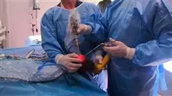
Veterinarians face new challenges every day in treating their patients. The Postgraduate certificate in Orthopedic Surgery in Large Animals comprises a complete and up-to-date educational program including the latest advances in traumatology and orthopedic surgery in ruminants (cattle, sheep), camelids (camels, alpacas and llamas), swine (pigs, wild boars) and equidae (horses, donkeys and mules).
The theoretical and practical content has been chosen taking into account its potential practical application in daily clinical practice. Furthermore, the audiovisual material collects scientific and practical information on the essential disciplines for professional practice.
In each topic, practical cases presented by experts in Traumatology and Orthopedic Surgery in Large Animals have been developed, with the objective of the practically applying the knowledge acquired. In addition, students will participate in a self-evaluation process to improve their learning and knowledge during their practical activities.
The teaching team of the Postgraduate certificate in Orthopedic Surgery in Large Animals has programmed a careful selection of techniques used in the diagnosis and treatment of ruminants (cattle, sheep), camelids (camels, alpacas, llamas), swine (pigs, wild boars) and equidae (horses, donkeys and mules), including the description of musculoskeletal surgery and rehabilitation in those species to which they are applied.
The teaching surgeons of this Postgraduate certificate are graduates of the European or American College of Veterinary Surgeons and have extensive experience both in the university field and in private practice. In both areas, they are responsible for large animal surgery services in leading veterinary centers and most of them direct residency programs, master's degree programs and research projects.
As a result of the training that the teaching staff of this Postgraduate certificate undertook in North America and Europe, the techniques have been extensively tested and are internationally recognized.
All of these elements mentioned above make this Postgraduate certificate a unique specialization program, exclusive and different to all the courses offered in other universities.
Do not miss the opportunity to take this Postgraduate certificate with TECH. It's the perfect opportunity to advance in your veterinary career"
This Postgraduate certificate in Orthopedic Surgery in Large Animals contains the most complete and up-to-date educational program on the market. The most important features include:
- Practical cases presented by experts in Advanced Techniques for Cardiovascular Pathology in Large Animals: Equidae, Ruminants and Swine
- The graphic, schematic, and practical contents with which they are created, provide scientific and practical information on the disciplines that are essential for professional development
- Latest innovations on Orthopedic Surgery in Large Animals
- Practical exercises where self-assessment can be used to improve learning
- Special emphasis on innovative methodologies in Orthopedic Surgery in Large Animals
- Theoretical lessons, questions to the expert, debate forums on controversial topics, and individual reflection assignments
- Content that is accessible from any fixed or portable device with an Internet connection
This course is the best investment you can make when choosing a refresher programme to update your existing knowledge of Large Animal Veterinary Medicine"
The multimedia content, developed with the latest educational technology, will provide the professional with situated and contextual learning, i.e., a simulated environment that will provide immersive learning programmed to train in real situations.
This program is designed around Problem-Based Learning, whereby the specialist must try to solve the different professional practice situations that arise throughout the program. For this purpose, the professional will be assisted by an innovative interactive video system created by renowned and experienced experts in Orthopedic Surgery in Large Animals.
This program comes with the best educational material, providing you with a contextual approach that will facilitate your learning"

This 100% online Postgraduate certificate will allow you to combine your studies with your professional work while increasing your knowledge in this field"
Why study at TECH?
TECH is the world’s largest online university. With an impressive catalog of more than 14,000 university programs available in 11 languages, it is positioned as a leader in employability, with a 99% job placement rate. In addition, it relies on an enormous faculty of more than 6,000 professors of the highest international renown.

Study at the world's largest online university and guarantee your professional success. The future starts at TECH”
The world’s best online university according to FORBES
The prestigious Forbes magazine, specialized in business and finance, has highlighted TECH as “the world's best online university” This is what they have recently stated in an article in their digital edition in which they echo the success story of this institution, “thanks to the academic offer it provides, the selection of its teaching staff, and an innovative learning method aimed at educating the professionals of the future”
A revolutionary study method, a cutting-edge faculty and a practical focus: the key to TECH's success.
The most complete study plans on the university scene
TECH offers the most complete study plans on the university scene, with syllabuses that cover fundamental concepts and, at the same time, the main scientific advances in their specific scientific areas. In addition, these programs are continuously being updated to guarantee students the academic vanguard and the most in-demand professional skills. In this way, the university's qualifications provide its graduates with a significant advantage to propel their careers to success.
TECH offers the most comprehensive and intensive study plans on the current university scene.
A world-class teaching staff
TECH's teaching staff is made up of more than 6,000 professors with the highest international recognition. Professors, researchers and top executives of multinational companies, including Isaiah Covington, performance coach of the Boston Celtics; Magda Romanska, principal investigator at Harvard MetaLAB; Ignacio Wistumba, chairman of the department of translational molecular pathology at MD Anderson Cancer Center; and D.W. Pine, creative director of TIME magazine, among others.
Internationally renowned experts, specialized in different branches of Health, Technology, Communication and Business, form part of the TECH faculty.
A unique learning method
TECH is the first university to use Relearning in all its programs. It is the best online learning methodology, accredited with international teaching quality certifications, provided by prestigious educational agencies. In addition, this disruptive educational model is complemented with the “Case Method”, thereby setting up a unique online teaching strategy. Innovative teaching resources are also implemented, including detailed videos, infographics and interactive summaries.
TECH combines Relearning and the Case Method in all its university programs to guarantee excellent theoretical and practical learning, studying whenever and wherever you want.
The world's largest online university
TECH is the world’s largest online university. We are the largest educational institution, with the best and widest online educational catalog, one hundred percent online and covering the vast majority of areas of knowledge. We offer a large selection of our own degrees and accredited online undergraduate and postgraduate degrees. In total, more than 14,000 university degrees, in eleven different languages, make us the largest educational largest in the world.
TECH has the world's most extensive catalog of academic and official programs, available in more than 11 languages.
Google Premier Partner
The American technology giant has awarded TECH the Google Google Premier Partner badge. This award, which is only available to 3% of the world's companies, highlights the efficient, flexible and tailored experience that this university provides to students. The recognition as a Google Premier Partner not only accredits the maximum rigor, performance and investment in TECH's digital infrastructures, but also places this university as one of the world's leading technology companies.
Google has positioned TECH in the top 3% of the world's most important technology companies by awarding it its Google Premier Partner badge.
The official online university of the NBA
TECH is the official online university of the NBA. Thanks to our agreement with the biggest league in basketball, we offer our students exclusive university programs, as well as a wide variety of educational resources focused on the business of the league and other areas of the sports industry. Each program is made up of a uniquely designed syllabus and features exceptional guest hosts: professionals with a distinguished sports background who will offer their expertise on the most relevant topics.
TECH has been selected by the NBA, the world's top basketball league, as its official online university.
The top-rated university by its students
Students have positioned TECH as the world's top-rated university on the main review websites, with a highest rating of 4.9 out of 5, obtained from more than 1,000 reviews. These results consolidate TECH as the benchmark university institution at an international level, reflecting the excellence and positive impact of its educational model.” reflecting the excellence and positive impact of its educational model.”
TECH is the world’s top-rated university by its students.
Leaders in employability
TECH has managed to become the leading university in employability. 99% of its students obtain jobs in the academic field they have studied, within one year of completing any of the university's programs. A similar number achieve immediate career enhancement. All this thanks to a study methodology that bases its effectiveness on the acquisition of practical skills, which are absolutely necessary for professional development.
99% of TECH graduates find a job within a year of completing their studies.
Postgraduate Certificate in Orthopedic Surgery in Large Animals
.
Are you a veterinarian looking to expand your skills in large animal orthopedic surgery? Are you interested in learning advanced surgical techniques and improving your knowledge in orthopedics? Then TECH Global University's Postgraduate Certificate in Orthopedic Surgery in Large Animals is for you. This program is designed to provide you with the skills and knowledge necessary to perform complex orthopedic surgeries on larger animals, such as horses and cattle. Through our team of orthopedic experts, you will learn advanced techniques for diagnosis, evaluation and treatment of orthopedic injuries, as well as pain management and postoperative rehabilitation.
Acquire the necessary knowledge to become a specialist in orthopedic surgeries in large animals.
The course is designed to enable you to adapt your skills to any clinical situation and improve your ability to perform orthopedic surgeries in the field of veterinary medicine. You will learn to perform surgical interventions from experts, and make critical decisions based on the analysis of data and information from each case. The program includes the opportunity to participate in real clinical cases and use state-of-the-art technology, which will allow you to gain practical and enriching experience in the field of veterinary orthopedics. Don't miss the opportunity to improve your skills in orthopedic surgeries in large animals. Join TECH Global University's Postgraduate Certificate in Orthopedic Surgery in Large Animals and become a specialist in the field of veterinary medicine!


