University certificate
The world's largest faculty of medicine”
Introduction to the Program
Master electroencephalograms and prove that you are a physician capable of meeting even greater healthcare challenges"

A medical professional who aspires to major professional improvements should look for a demanded and current specialization, with which to stand out from his or her colleagues. Clinical neurophysiology, and more specifically electroencephalograms, are often overlooked when looking for a particular specialty given their common use for the diagnosis of various pathologies.
But this is precisely its strong and attractive point for the medical professional who wants to stand out, because having a full understanding of the most intrinsic and detailed aspects of encephalograms, they will quickly become an essential part of the healthcare organization chart in which they find themselves.
This TECH Postgraduate certificate brings together, therefore, an extensive and complete syllabus ranging from standard protocols and maneuvers for performing EGG to slow and epileptiform anomalies that the professional may encounter. It also focuses on quantified EGG, a current method that requires state-of-the-art software to see the dynamic changes that occur during cognitive processing tasks, giving the clinician the ability to identify which areas of the brain may be compromised and which are functioning properly.
A completely online program that adapts to the needs of its students, giving them the possibility of taking it completely at their own pace and specific needs. The student has access to all the didactic material from the first day of the Postgraduate certificate, being able to download it to any device with internet access.
You will be prepared to know how to recognize any abnormalities in the EEGs you perform, which will make you vital to your healthcare team"
This Postgraduate certificate in Brain Electrogenesis. Recording and Analysis Techniques. Electroencephalogram Development contains the most complete and up to date educational program on the market. The most important features include:
- The development of case studies presented by medical experts in neurophysiology and electroencephalograms
- The graphic, schematic, and eminently practical contents with which they are created, provide scientific and practical information on the disciplines that are essential for professional practice
- Practical exercises where self-assessment can be used to improve learning
- Its special emphasis on innovative methodologies
- Theoretical lessons, questions to the expert, debate forums on controversial topics, and individual reflection assignments
- Content that is accessible from any fixed or portable device with an Internet connection
Your own staff will benefit from having you as a reference when performing EGG on all kinds of patients"
The program’s teaching staff includes professionals from the sector who contribute their work experience to this training program, as well as renowned specialists from leading societies and prestigious universities.
The multimedia content, developed with the latest educational technology, will provide the professional with situated and contextual learning, i.e., a simulated environment that will provide immersive training programmed to train in real situations.
The design of this Program focuses on Problem-Based Learning, by means of which the professional will have to try to solve the different situations of Professional Practice, which will be posed throughout the Program. For this purpose, the student will be assisted by an innovative interactive video system created by renowned and experienced experts.
You have in your hands the possibility of specializing in a unique and distinctive field in the healthcare field. Don't miss it and enroll now"

y adding this Postgraduate certificate to your resume, you will have more chances to move up the career ladder and gain access to more prestigious healthcare positions"
Why study at TECH?
TECH is the world’s largest online university. With an impressive catalog of more than 14,000 university programs available in 11 languages, it is positioned as a leader in employability, with a 99% job placement rate. In addition, it relies on an enormous faculty of more than 6,000 professors of the highest international renown.

Study at the world's largest online university and guarantee your professional success. The future starts at TECH”
The world’s best online university according to FORBES
The prestigious Forbes magazine, specialized in business and finance, has highlighted TECH as “the world's best online university” This is what they have recently stated in an article in their digital edition in which they echo the success story of this institution, “thanks to the academic offer it provides, the selection of its teaching staff, and an innovative learning method aimed at educating the professionals of the future”
A revolutionary study method, a cutting-edge faculty and a practical focus: the key to TECH's success.
The most complete study plans on the university scene
TECH offers the most complete study plans on the university scene, with syllabuses that cover fundamental concepts and, at the same time, the main scientific advances in their specific scientific areas. In addition, these programs are continuously being updated to guarantee students the academic vanguard and the most in-demand professional skills. In this way, the university's qualifications provide its graduates with a significant advantage to propel their careers to success.
TECH offers the most comprehensive and intensive study plans on the current university scene.
A world-class teaching staff
TECH's teaching staff is made up of more than 6,000 professors with the highest international recognition. Professors, researchers and top executives of multinational companies, including Isaiah Covington, performance coach of the Boston Celtics; Magda Romanska, principal investigator at Harvard MetaLAB; Ignacio Wistumba, chairman of the department of translational molecular pathology at MD Anderson Cancer Center; and D.W. Pine, creative director of TIME magazine, among others.
Internationally renowned experts, specialized in different branches of Health, Technology, Communication and Business, form part of the TECH faculty.
A unique learning method
TECH is the first university to use Relearning in all its programs. It is the best online learning methodology, accredited with international teaching quality certifications, provided by prestigious educational agencies. In addition, this disruptive educational model is complemented with the “Case Method”, thereby setting up a unique online teaching strategy. Innovative teaching resources are also implemented, including detailed videos, infographics and interactive summaries.
TECH combines Relearning and the Case Method in all its university programs to guarantee excellent theoretical and practical learning, studying whenever and wherever you want.
The world's largest online university
TECH is the world’s largest online university. We are the largest educational institution, with the best and widest online educational catalog, one hundred percent online and covering the vast majority of areas of knowledge. We offer a large selection of our own degrees and accredited online undergraduate and postgraduate degrees. In total, more than 14,000 university degrees, in eleven different languages, make us the largest educational largest in the world.
TECH has the world's most extensive catalog of academic and official programs, available in more than 11 languages.
Google Premier Partner
The American technology giant has awarded TECH the Google Google Premier Partner badge. This award, which is only available to 3% of the world's companies, highlights the efficient, flexible and tailored experience that this university provides to students. The recognition as a Google Premier Partner not only accredits the maximum rigor, performance and investment in TECH's digital infrastructures, but also places this university as one of the world's leading technology companies.
Google has positioned TECH in the top 3% of the world's most important technology companies by awarding it its Google Premier Partner badge.
The official online university of the NBA
TECH is the official online university of the NBA. Thanks to our agreement with the biggest league in basketball, we offer our students exclusive university programs, as well as a wide variety of educational resources focused on the business of the league and other areas of the sports industry. Each program is made up of a uniquely designed syllabus and features exceptional guest hosts: professionals with a distinguished sports background who will offer their expertise on the most relevant topics.
TECH has been selected by the NBA, the world's top basketball league, as its official online university.
The top-rated university by its students
Students have positioned TECH as the world's top-rated university on the main review websites, with a highest rating of 4.9 out of 5, obtained from more than 1,000 reviews. These results consolidate TECH as the benchmark university institution at an international level, reflecting the excellence and positive impact of its educational model.” reflecting the excellence and positive impact of its educational model.”
TECH is the world’s top-rated university by its students.
Leaders in employability
TECH has managed to become the leading university in employability. 99% of its students obtain jobs in the academic field they have studied, within one year of completing any of the university's programs. A similar number achieve immediate career enhancement. All this thanks to a study methodology that bases its effectiveness on the acquisition of practical skills, which are absolutely necessary for professional development.
99% of TECH graduates find a job within a year of completing their studies.
Postgraduate Certificate in Brain Electrogenesis. Recording and Analysis Techniques. Development of the Electroencephalogram.
Brain electrogenesis is the process by which brain cells, called neurons, generate a small electric field due to the communication and transmission of information. The technique of recording and analyzing brain electrogenesis is known as electroencephalography (EEG).
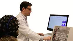
EEG is a noninvasive technique in which electrodes are placed on the scalp to record the electrical activity of the brain. The activity is recorded as a pattern of waves of different frequencies and amplitudes. EEG can be used to detect and diagnose a variety of neurological conditions, including seizure disorders, neurodegenerative diseases, and sleep disorders, among others.
The development of the electroencephalogram (EEG) began in the late 19th century, when neuropsychiatrist Hans Berger made the first recordings of electrical activity in the human brain using electrodes attached to the scalp. After years of research, in 1929 Berger published the first long-term EEG recording and established the use of the technique for investigating brain activity.
Since then, EEG has steadily evolved with the introduction of more advanced technologies and techniques. Current EEG technology can record brain activity in real time and in high spatial and temporal resolution, allowing for a deeper understanding of normal and abnormal brain activity.
In conclusion brain electrogenesis is the process by which brain cells generate an electrical field that can be recorded using the electroencephalography (EEG) technique. The technique is used to detect and diagnose neurological conditions and has evolved steadily since its discovery in the 19th century.
During the Postgraduate Certificate course, students will learn the theoretical basics of brain electrophysiology and the generation of electrical signals in the brain. In addition, they will learn how to use the technique of EEG recording and analysis, through procedures of placing electrodes on the scalp, understanding the reading and analysis of the recorded brain signals.


