University certificate
The world's largest faculty of medicine”
Introduction to the Program
You will master digital image processing thanks to the best digital university in the world, according to Forbes"

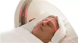
The Compton Effect is one of the most important processes to keep in mind when calculating radiation dose in treatments. The reasons lie in the implications it has on the generation of medical images and radiation dosage in different therapies. If experts were to make mistakes when measuring this process, this would lead to everything from incorrect diagnoses to radiation overdosage. This, in turn, could lead to side effects and damage to normal tissues.
In order to obtain proper education on fabric composition and density, TECH has implemented this advanced Postgraduate diploma. In this way, nurses will be able to carry out safe clinical practices, using both X-Ray and Gamma Rays. In fact, the curriculum will address the interactions between photons and matter.
It will also delve into the weighting factors of organs according to their radiosensitivity, analyzing various tools for quality control in visualization systems. This will allow the graduate to identify the risks in the hospital area and to design structural shielding for the protection of both patients and personnel.
In order to consolidate these contents, the methodology of this program reinforces its innovative character. In this way, TECH offer a 100% online educational environment. to the needs of busy professionals looking to advance their careers. In addition, it will employ the Relearning methodology, based on the repetition of key concepts to fix knowledge and facilitate learning. In this way, the combination of flexibility and a robust pedagogical approach makes it highly accessible. In addition, learners will have access to an extensive library of innovative multimedia resources in different audiovisual formats, such as interactive summaries, explanatory videos, photographs, case studies and infographics.
You will delve into the interaction between photons and matter to irradiate tumors with high precision"
This Postgraduate diploma in Radiophysics Applied to Diagnostic Imaging contains the most complete and up-to-date scientific program on the market. The most important features include:
- The development of case studies presented by experts in Radiophysics applied to Diagnostic Imaging
- The graphic, schematic and practical contents with which it is conceived gather scientific and practical information on those disciplines that are essential for professional practice
- Practical exercises where the self-assessment process can be carried out to improve learning
- Its special emphasis on innovative methodologies
- Theoretical lessons, questions to the expert, debate forums on controversial topics, and individual reflection assignments
- The availability of access to content from any fixed or portable device with an Internet connection
Looking to get the most out of Mammography equipment? Develop the most advanced tests in quality control, thanks to TECH"
The program’s teaching staff includes professionals from the sector who contribute their work experience to this program, as well as renowned specialists from leading societies and prestigious universities.
The multimedia content, developed with the latest educational technology, will provide the professional with situated and contextual learning, i.e., a simulated environment that will provide immersive education programmed to learn in real situations.
This program is designed around Problem-Based Learning, whereby the professional must try to solve the different professional practice situations that arise during the academic year For this purpose, the students will be assisted by an innovative interactive video system created by renowned and experienced experts.
You will cover dosimeter calibration in detail to ensure reliable radiation exposure measurements"

With the Relearning system, pioneer in TECH, you will reduce long hours of study and memorization"
Why study at TECH?
TECH is the world’s largest online university. With an impressive catalog of more than 14,000 university programs available in 11 languages, it is positioned as a leader in employability, with a 99% job placement rate. In addition, it relies on an enormous faculty of more than 6,000 professors of the highest international renown.

Study at the world's largest online university and guarantee your professional success. The future starts at TECH”
The world’s best online university according to FORBES
The prestigious Forbes magazine, specialized in business and finance, has highlighted TECH as “the world's best online university” This is what they have recently stated in an article in their digital edition in which they echo the success story of this institution, “thanks to the academic offer it provides, the selection of its teaching staff, and an innovative learning method aimed at educating the professionals of the future”
A revolutionary study method, a cutting-edge faculty and a practical focus: the key to TECH's success.
The most complete study plans on the university scene
TECH offers the most complete study plans on the university scene, with syllabuses that cover fundamental concepts and, at the same time, the main scientific advances in their specific scientific areas. In addition, these programs are continuously being updated to guarantee students the academic vanguard and the most in-demand professional skills. In this way, the university's qualifications provide its graduates with a significant advantage to propel their careers to success.
TECH offers the most comprehensive and intensive study plans on the current university scene.
A world-class teaching staff
TECH's teaching staff is made up of more than 6,000 professors with the highest international recognition. Professors, researchers and top executives of multinational companies, including Isaiah Covington, performance coach of the Boston Celtics; Magda Romanska, principal investigator at Harvard MetaLAB; Ignacio Wistumba, chairman of the department of translational molecular pathology at MD Anderson Cancer Center; and D.W. Pine, creative director of TIME magazine, among others.
Internationally renowned experts, specialized in different branches of Health, Technology, Communication and Business, form part of the TECH faculty.
A unique learning method
TECH is the first university to use Relearning in all its programs. It is the best online learning methodology, accredited with international teaching quality certifications, provided by prestigious educational agencies. In addition, this disruptive educational model is complemented with the “Case Method”, thereby setting up a unique online teaching strategy. Innovative teaching resources are also implemented, including detailed videos, infographics and interactive summaries.
TECH combines Relearning and the Case Method in all its university programs to guarantee excellent theoretical and practical learning, studying whenever and wherever you want.
The world's largest online university
TECH is the world’s largest online university. We are the largest educational institution, with the best and widest online educational catalog, one hundred percent online and covering the vast majority of areas of knowledge. We offer a large selection of our own degrees and accredited online undergraduate and postgraduate degrees. In total, more than 14,000 university degrees, in eleven different languages, make us the largest educational largest in the world.
TECH has the world's most extensive catalog of academic and official programs, available in more than 11 languages.
Google Premier Partner
The American technology giant has awarded TECH the Google Google Premier Partner badge. This award, which is only available to 3% of the world's companies, highlights the efficient, flexible and tailored experience that this university provides to students. The recognition as a Google Premier Partner not only accredits the maximum rigor, performance and investment in TECH's digital infrastructures, but also places this university as one of the world's leading technology companies.
Google has positioned TECH in the top 3% of the world's most important technology companies by awarding it its Google Premier Partner badge.
The official online university of the NBA
TECH is the official online university of the NBA. Thanks to our agreement with the biggest league in basketball, we offer our students exclusive university programs, as well as a wide variety of educational resources focused on the business of the league and other areas of the sports industry. Each program is made up of a uniquely designed syllabus and features exceptional guest hosts: professionals with a distinguished sports background who will offer their expertise on the most relevant topics.
TECH has been selected by the NBA, the world's top basketball league, as its official online university.
The top-rated university by its students
Students have positioned TECH as the world's top-rated university on the main review websites, with a highest rating of 4.9 out of 5, obtained from more than 1,000 reviews. These results consolidate TECH as the benchmark university institution at an international level, reflecting the excellence and positive impact of its educational model.” reflecting the excellence and positive impact of its educational model.”
TECH is the world’s top-rated university by its students.
Leaders in employability
TECH has managed to become the leading university in employability. 99% of its students obtain jobs in the academic field they have studied, within one year of completing any of the university's programs. A similar number achieve immediate career enhancement. All this thanks to a study methodology that bases its effectiveness on the acquisition of practical skills, which are absolutely necessary for professional development.
99% of TECH graduates find a job within a year of completing their studies.
Postgraduate Diploma in Radiophysics Applied to Diagnostic Imaging
Radiophysics applied to diagnostic imaging focuses on the application of physical principles and advanced techniques to ensure quality and safety in diagnostic imaging procedures. Would you like to specialize in this innovative field? TECH Global University has the ideal option for you: the Postgraduate Diploma in Radiophysics Applied to Diagnostic Imaging. This online program will provide you with an in-depth understanding of the physical principles that drive diagnostic imaging technologies. You will delve into the physical principles that govern the formation of medical images using various technologies, from radiography to magnetic resonance imaging. In addition, you will learn about the most advanced equipment and technologies used in diagnostic imaging. From computed tomography (CT), to ultrasound and magnetic resonance imaging systems, you will be guided through the unique characteristics of each modality and their application in clinical practice.
Learn about radiophysics applied to diagnostic imaging
Only at TECH will you find the most up-to-date methods in the field, complemented by multimedia material and completely new dynamic classes. As you advance through the training, you will be immersed in image quality and dosimetry optimization. You will explore techniques and practices to ensure high-quality diagnostic images while minimizing radiation exposure, ensuring safe and accurate procedures. In addition, you will understand the principles of dosimetry specific to diagnostic radiology, gaining tools to measure and calculate the radiation dose delivered during diagnostic procedures, contributing to the safety and efficacy of studies. Finally, you will explore both quality control and radiation safety in diagnostic imaging, as well as technological developments and current trends in the field of radiophysics applied to diagnostic imaging. Upon completion, you will be ready to lead the field of diagnostic imaging. This program will equip you with advanced knowledge and specialized skills to contribute to the advancement and excellence in the science behind medical imaging. Enroll now and take your career to the next level!


