
University certificate


The world's largest faculty of veterinary medicine”
Introduction to the Program
Esta especialización es la mejor opción que podrás encontrar para capacitarte y realizar diagnósticos más precisos”
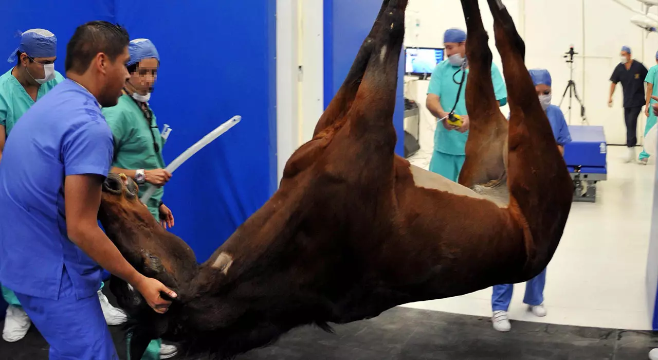
Los veterinarios se enfrentan cada día a nuevos retos para tratar a sus pacientes. La Postgraduate diploma en Orthopedic Surgery in Large Animals: Ruminants, Camelids, Swine and Equidae comprende un programa educativo completo y actualizado incluyendo los últimos avances en Traumatología y Cirugía Ortopédica en Rumiantes (Bovino, Ovino), Camélidos (Camellos, Alpacas y Llamas) Suidos (Cerdos, Jabalíes) y Équidos (Caballos, Burros y Mulas).
El contenido teórico-práctico ha sido seleccionado teniendo en cuenta su potencial de aplicación práctica en la clínica diaria. Además, el material audiovisual recoge una información científica y práctica sobre las disciplinas indispensables para el ejercicio profesional.
En cada tema se han desarrollado casos prácticos presentados por expertos en Traumatología y Cirugía Ortopédica en Especies Mayores, teniendo como objetivo la aplicación práctica de los conocimientos adquiridos. Además, el alumno en sus actividades prácticas participará en un proceso de autoevaluación para mejorar el aprendizaje y sus conocimientos.
El equipo docente de la Postgraduate diploma en Orthopedic Surgery in Large Animals: Ruminants, Camelids, Swine and Equidae ha programado una cuidadosa selección de técnicas usadas en el diagnóstico y tratamiento de Cojeras en Rumiantes (Bovino, Ovino), Camélidos (Camellos, Alpacas, Llamas), Suidos (Cerdos, Jabalíes) y Équidos (Caballos, Burros y Mulas), incluyendo la descripción de las intervenciones quirúrgicas musculoesqueléticas y de rehabilitación en aquellas especies en que se practiquen.
Los cirujanos profesores de esta Postgraduate diploma son Diplomados por el Colegio Europeo o Americano de Cirujanos Veterinarios y poseen una amplia experiencia tanto en ámbito universitario como en clínica privada. En ambos ámbitos son responsables de los servicios de cirugía de Especies Mayores en centros veterinarios de referencia y la mayoría de ellos dirigen programas de residencia, máster y proyectos de investigación.
Como consecuencia de la capacitación del profesorado de esta Postgraduate diploma en América del Norte y Europa, las técnicas desarrolladas han sido ampliamente contrastadas y son reconocidas internacionalmente.
No dejes pasar la ocasión de realizar esta Postgraduate diploma con TECH. Es la oportunidad perfecta para avanzar en tu carrera veterinaria”
Esta Postgraduate diploma en Orthopedic Surgery in Large Animals: Ruminants, Camelids, Swine and Equidae contiene el programa educativo más completo y actualizado del mercado. Las características más destacadas de la capacitación son:
- El desarrollo de casos prácticos presentados por expertos en Cirugía Ortopédica en Especies Mayores: Rumiantes, Camélidos, Suidos y Équidos
- Los contenidos gráficos, esquemáticos y eminentemente prácticos con los que están concebidos recogen una información científica y práctica sobre aquellas disciplinas indispensables para el ejercicio profesional
- Las novedades sobre Cirugía Ortopédica en Especies Mayores: Rumiantes, Camélidos, Suidos y Équidos
- Los ejercicios prácticos donde realizar el proceso de autoevaluación para mejorar el aprendizaje
- Su especial hincapié en metodologías innovadoras en Cirugía Ortopédica en Especies Mayores: Rumiantes, Camélidos, Suidos y Équidos
- Las lecciones teóricas, preguntas al experto, foros de discusión de temas controvertidos y trabajos de reflexión individual
- La disponibilidad de acceso a los contenidos desde cualquier dispositivo fijo o portátil con conexión a internet
Analizarás las complicaciones anestésicas más frecuentes en la clínica de Especies Mayores, y, en particular, en referencia a la cirugía ortopédica”
Su contenido multimedia, elaborado con la última tecnología educativa, permitirá al profesional un aprendizaje situado y contextual, es decir, un entorno simulado que proporcionará una capacitación inmersiva programada para entrenarse ante situaciones reales.
El diseño de este programa se centra en el Aprendizaje Basado en Problemas, mediante el cual el especialista deberá tratar de resolver las distintas situaciones de práctica profesional que se le planteen a lo largo del curso académico. Para ello, el profesional contará con la ayuda de un novedoso sistema de vídeo interactivo realizado por reconocidos expertos en Cirugía Ortopédica en Especies Mayores: Rumiantes, Camélidos, Suidos y Équidos y con gran experiencia.
Esta capacitación cuenta con el mejor material didáctico, lo que te permitirá un estudio contextual que te facilitará el aprendizaje”
Los veterinarios deben continuar su especialización para adaptarse a los nuevos avances en este campo"
Syllabus
The structure of the contents has been designed by the best professionals in the field of Orthopedic Surgery in Large Animals: Ruminants, Camelids, Swine and Equidae with extensive experience and recognized prestige in the profession, backed by the volume of cases reviewed, studied, and diagnosed, and with extensive knowledge of new technologies applied to veterinary medicine.

This Postgraduate diploma in Orthopedic Surgery in Large Animals: Ruminants, Camelids, Swine and Equidae contains the most complete and up-to-date scientific program on the market”
Module 1. Preoperative Aspects in Large Animals: Ruminants, Swine and Equidae
1.1. Preparation for Surgery: Decision Making, Operation Risks, Patient Considerations
1.1.1. Surgical Risk
1.1.2. Preoperative Patient Evaluation
1.2. Pharmacological Management for On-Site Procedures
1.2.1. Sedation Drugs
1.2.2. Continuous Infusions
1.2.3. Local Anesthetics
1.2.4. Containment Systems, Other Considerations
1.2.5. Selection of Procedures to be Performed On Site
1.3. General Anesthesia
1.3.1. Inhalation General Anesthesia
1.3.2. Intravenous General Anesthesia
1.4. Recovery from General Anesthesia
1.4.1. Management During Recovery
1.4.2. Factors Affecting Recovery
1.4.3. Different Techniques or Installations for Anesthetic Recovery
1.5. General Surgical Technique
1.5.1. General Aspects
1.5.2. Basic Manipulation of Surgical Instruments
1.5.3. Tissue Incision, Blunt Dissection
1.5.4. Tissue Retraction and Handling
1.5.5. Surgical Irrigation and Suction
1.6. Preparation of the Surgery, Personnel, Patient and Surgical Area
1.6.1. Pre-surgery Planning
1.6.2. Surgical Attire, Preparation of Surgical Equipment: Gloves, Gowns etc.
1.6.3. Preparation of the Patient and Surgical Area
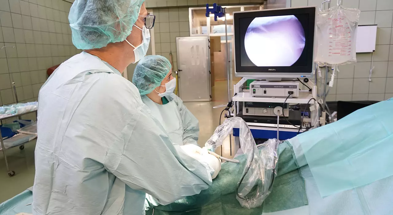
1.7. Use of Diagnostic Imaging in Orthopedic Surgery
1.7.1. Diagnostic Imaging Techniques
1.7.2. Diagnostic Imaging in Preparation for Surgery
1.7.3. Use of the Intraoperation Image
1.8. Disinfection of Material, Sterilization
1.8.1. Cold Disinfection
1.8.2. Packaging the Material
1.8.3. Different Autoclaves and Sterilizing Products
1.9. Orthopedic Surgical Instruments in Large Animals
1.9.1. General Instruments in Orthopedics
1.9.2. Arthroscopic Instruments
1.9.3. Osteosynthesis Instruments
1.10. The Operating Room for Large Animals
1.10.1. Basic Installations
1.10.2. Importance of the Design of the Operating Room, Asepsis
1.10.3. Technical Specifications of the Advanced Surgical Equipment
Module 2. Reparation of Fractures in Large Animals Ruminants, Swine and Equidae
2.1. Bone Metabolism and Healing
2.1.1. Anatomy
2.1.2. Histological Structure
2.1.3. Bone Healing
2.1.4. Biomechanics of the Bone
2.1.5. Classification of Fractures
2.2. Stabilization of Fractures in an Emergency, Decision Making and Transport
2.2.1. Clinical Examination of a Patient With a Suspected Fracture
2.2.2. Stabilization of a Patient With Fractures
2.2.3. Transport of a Patient With a Fracture
2.2.4. Stabilization of Fractures, Decision Making and Transport of Ruminants (Cattle, Sheep), Camelids (Camels, Alpacas and Llamas) and Swine (Pigs, Wild Boar)
2.3. External Coaptation
2.3.1. Placement of Robert Jones Bandages
2.3.2. Placement of Acrylic Casts
2.3.3. Splints, Bandages With Casts and Combinations
2.3.4. Complications of Acrylic Casts
2.3.5. Removal of Acrylic Casts
2.4. Reducing Fractures, Management of Soft Tissue in the Approach
2.4.1. Displacements of Fracture Strands
2.4.2. Objectives of the Fracture Reduction
2.4.3. Reduction Techniques
2.4.4. Evaluation of Reduction
2.4.5. Management of Soft Tissues
2.4.6. Histology and Blood Supply of the Skin
2.4.7. Physical Properties and Biomechanics of the Skin
2.4.8. Planning the Approach
2.4.9. Incisions
2.4.10. Wound Closure
2.5. Materials for Implants in Large Animals
2.5.1. Properties of the Materials
2.5.2. Stainless Steel
2.5.3. Titanium
2.5.4. Material Fatigue
2.6. External Fixators
2.6.1. Transfixion Casts
2.6.2. External Fixators
2.6.3. External Fixators of Ruminants (Cattle, Sheep), Camelids (Camels, Alpacas and Llamas) and Swine (Pigs, Wild Boar)
2.7. Instruments for Inserting an Implant
2.7.1. Plate Contouring Instruments
2.7.2. Instruments for Inserting Screws
2.7.3. Instruments for Inserting Plates
2.8. Implants
2.8.1. Screws
2.8.2. Plates
2.8.3. Placement Techniques
2.8.4. Functions of Each Implant
2.8.5. Tension Band
2.9. Bone Grafts
2.9.1. Indications
2.9.2. Removal Sites
2.9.3. Complications
2.9.4. Synthetic Bone Grafts
2.10. Complications of Inserting an Implant
2.10.1. Lack of Reduction
2.10.2. Incorrect Number and Size of Implants
2.10.3. Incorect Position of the Implant
2.10.4. Complications Related to the Compression Screw
2.10.5. Complications Related to Plates
Module 3. Common Orthopedic Surgery Procedures of the Musculoskeletal System in Large Animals: Ruminants, Swine and Equidae Part I
3.1. Fractures of Distal Phalanx and Navicular Bone
3.1.1. Distal Phalanx
3.1.1.1. Causes
3.1.1.2. Classification
3.1.1.3. Clinical Signs
3.1.1.4. Treatment
3.1.2. Navicular Bone Fracture
3.1.2.1. Causes
3.1.2.2. Clinical Signs and Diagnosis
3.1.2.3. Treatment
3.1.3. Digital Neurectomy
3.1.4. Bovine Distal Phalanx Fracture
3.1.5. Bovine Pedal Osteitis
3.1.6. Sepsis of the Common Digital Flexor Tendon Sheath in Ruminants
3.1.6.1. Tenosynoviotomy With Resection of Affected Tissue
3.2. Middle Phalanx Fracture
3.2.1. Etiology
3.2.2. Clinical Signs
3.2.3. Diagnosis
3.2.4. Settings
3.2.4.1. Palmar/Plantar Eminence Fractures
3.2.4.1.1. Uni- and Biaxial Fractures
3.2.4.2. Axial Fractures
3.2.4.3. Comminuted Fractures
3.3. Proximal Phalangeal and Proximal Interphalangeal Joints
3.3.1. Osteoarthritis
3.3.2. Subchondral Cystic Lesions
3.3.3. Dislocations and Subluxations
3.3.4. Fracture Configurations
3.3.5. Clinical Signs
3.3.6. Diaphyseal Fractures
3.3.7. Incomplete Sagittal Fractures
3.3.8. Non-Displaced Long Incomplete Sagittal Incomplete Fractures
3.3.9. Displaced Complete Sagittal Fractures
3.3.10. Frontal Fractures
3.3.11. Comminuted Fractures
3.4. Metacarpal- Metatarsal Falangeal Joint
3.4.1. Proximal Sesamoid Bone Fractures
3.4.1.1. Mid-Body
3.4.1.2. Basal
3.4.1.3. Abaxial
3.4.1.4. Sagittal
3.4.1.5. Biaxial
3.4.2. Osteoarthritis
3.4.3. Subchondral Cystic Lesions
3.4.4. Dislocation
3.4.5. Tenosynovitis/Desmitis/Constriction of the Annular Ligament
3.4.5.1. Mass Removal
3.4.5.1. Section of the Annular Ligament
3.4.5.1. Tendon Debridement
3.5. Metacarpal/Metatarsal Bones
3.5.1. Lateral Condylar Fractures
3.5.1.1. Signs
3.5.1.2. Diagnosis
3.5.1.3. Emergency Treatment
3.5.1.4. Surgery of Displaced Fractures
3.5.1.5. Surgery of Non-Displaced Fractures
3.5.2. Medial Condylar Fractures
3.5.2.1. Open Approach Surgery
3.5.2.2. Minimally Invasive Surgery
3.5.2.3. Post-Operative Care
3.5.2.4. Prognosis
3.5.3. Transverse Fractures of the Distal Diaphysis of the Third Metacarpal Bone
3.5.3.1. Non-Surgical Treatment
3.5.3.2. Surgical Treatment
3.5.3.3. Prognosis
3.5.4. Diaphyseal Fractures
3.5.4.1. Non-Surgical Management
3.5.4.2. Surgical Management
3.5.4.3. Prognosis
3.5.5. Distal Physial Fractures
3.5.6. Proximal Articular Fractures
3.5.7. Dorsal Cortical Fractures
3.5.7.1. Non-Surgical Management
3.5.7.2. Surgical Treatment
3.5.7.3. Prognosis
3.5.8. Metacarpal/Metatarsal Bone Fractures in Ruminants (Cattle, Sheep) and Camelids (Camels, Alpacas and Llamas)
3.6. Rudimentary Metacarpal/Metatarsal Bones
3.6.1. Fractures
3.6.2. Clinical Examination
3.6.3. Diagnosis
3.6.4. Proximal Fractures
3.6.4.1. Debridement
3.6.4.2. Internal Fixation
3.6.4.3. Ostectomy
3.6.4.4. Complete Removal
3.6.4.5. Prognosis
3.6.4.6. Complications
3.6.5. Mid-Body Fractures
3.6.5.1. Non-Surgical Management
3.6.5.2. Surgical Treatment
3.6.5.3. Prognosis
3.6.6. Distal Fractures
3.6.6.1. Non-Surgical Management
3.6.6.2. Surgical Treatment
3.6.6.3. Prognosis
3.6.7. Exostosis
3.6.7.1. Pathophysiology
3.6.7.2. Clinical Examination
3.6.7.3. Diagnosis
3.6.7.3.1. Treatment
3.6.7.3.2. Non-Surgical Management
3.6.7.3.3. Surgical Management
3.6.7.4. Prognosis
3.6.8. Polydactyly in Ruminants and Equidae
3.6.9. Neoplasty
3.7. Tendon and Ligament Pathologies That Can Be Resolved Surgically
3.7.1. Carporadic Extensor Carpi Radialis Tendon Rupture
3.7.1.1. Pathophysiology
3.7.1.2. Diagnosis
3.7.1.3. Treatment
3.7.1.4. Prognosis
3.7.2. Biceps Brachii Tendon and Infraspinatus Tendon Pathologies
3.7.2.1. Treatment
3.7.2.1.1. Biceps Tendon Transection
3.7.2.2. Prognosis
3.7.3. Surgery for Suspensory Ligament Desmopathy in the Forelimb
3.7.4. Surgery of Suspensory Ligament Branches
3.7.5. Suspensory Ligament Damage in Ruminants
3.7.6. Tenectomy of the Medial Head of the Deep Digital Flexor Tendon
3.7.7. Surgery for Suspensory Ligament Dismopathy of the Hind Limb
3.7.8. Intermittent Patella Fixation in Equidae
3.7.9. Patella Fixation in Ruminants
3.7.10. Tears or Avulsions of Collateral Ligaments in Ruminants
3.7.11. Cranial Cruciate Ligament Rupture in Ruminants
3.7.11.1. Peri-Surgical Planning
3.7.11.2. Imbrication of Stifle Joint
3.7.11.3. Cranial Cruciate Ligament Replacement
3.7.11.3.1. With Gluteobiceps Tendon
3.7.11.3.2. With Synthetic Material
3.7.11.3.3. Post-Surgery and Prognosis
3.7.12. Damage to Collateral Ligaments of the Stifle
3.7.12.1. Surgery
3.7.12.2. Prognosis
3.7.13. Superficial Digital Flexor Tendon Dislocation
3.8. Muscle Pathologies That Can Be Resolved Surgically
3.8.1. Fibrotic Myopathy
3.8.1.1. Pathophysiology
3.8.1.2. Diagnosis
3.8.1.3. Treatment
3.8.1.4. Prognosis
3.8.2. Arpeo (Equine Reflex Hypertonia)
3.8.2.1. Pathophysiology
3.8.2.2. Diagnosis
3.8.2.3. Treatment
3.8.2.4. Prognosis
3.8.3. Third Peroneal
3.8.3.1. Pathophysiology
3.8.3.2. Diagnosis
3.8.3.3. Treatment
3.8.3.4. Prognosis
3.8.4. Rupture and Avulsion of the Gastrocnemius Muscles
3.8.4.1. Pathophysiology
3.8.4.2. Diagnosis
3.8.4.3. Treatment
3.8.4.4. Prognosis
3.8.5. Aerophagia
3.8.5.1. Pathophysiology
3.8.5.2. Diagnosis
3.8.5.3. Treatment
3.8.5.4. Prognosis
3.8.6. Spastic Paresis
3.9. Arthrodesis
3.9.1. Equine Distal Interphalangeal Joint
3.9.2. Arthrodesis of the Distal Bovine Interphalangeal Joint
3.9.3. Proximal Interphalangeal Joint
3.9.4. Metacarpal/Metatarsophalangeal Joint
3.9.5. Of the Carpus
3.9.6. Of the Shoulder
3.9.7. Of Distal Tarsal Joints
3.9.8. Talocalcaneal
3.10. Laminitis and Amputations in Ruminants, Swine and Equidae
3.10.1. Laminitis
3.10.1.1. Deep Digital Flexor Tendon Tenotomy
3.10.1.1.1. At Pastern Level
3.10.1.1.2. At Mid Metacarpal-Metatarsal Level
3.10.1.2. Prognosis
3.10.2. Amputations in Ruminants, Swine and Equidae
3.10.2.1. Bovine Digit Amputation
3.10.2.2. Bovine Extra Digit Amputation
3.10.2.3. Tail Amputation
3.10.2.4. Limb Amputation
3.10.2.5. Specifics in Swine
Module 4. Common Orthopedic Surgery Procedures of the Musculoskeletal System in Large Animals: Ruminants, Swine and Equidae Part II
4.1. Carpus
4.1.1. Pathophysiology
4.1.2. Multifragmentary Fractures
4.1.2.1. Pathogenesis
4.1.2.2. Diagnosis
4.1.2.3. Treatment
4.1.3. Accessory Bone Fracture
4.1.3.1. Pathogenesis
4.1.3.2. Diagnosis
4.1.3.3. Treatment
4.1.3.4. Non-Surgical Management
4.1.3.5. Surgical Management
4.1.3.6. Prognosis
4.1.4. Carpal Hygroma
4.1.5. Radial Distal Exostosis
4.1.5.1. Clinical Examination
4.1.5.2. Diagnosis
4.1.5.3. Treatment
4.1.5.3.1. Non-Surgical Management
4.1.5.3.2. Surgical Management
4.1.5.4. Prognosis
4.1.6. Dislocation
4.1.6.1. Pathogenesis
4.1.6.2. Diagnosis
4.1.6.3. Treatment
4.1.6.3.1. Non-Surgical Management
4.1.6.3.2. Surgical Management
4.1.6.4. Prognosis
4.1.7. Coronation
4.1.7.1. Pathogenesis
4.1.7.2. Diagnosis
4.1.7.3. Treatment
4.1.8. Synovial Osteochondromatosis
4.1.9. Circumscribed Calcinosis
4.1.9.1. Pathophysiology
4.1.9.2. Diagnosis
4.1.9.3. Treatment
4.1.9.4. Prognosis
4.2. Radio and Ulna
4.2.1. Ulna Fracture
4.2.1.1. Anatomy
4.2.1.2. Pathogenesis
4.2.1.3. Diagnosis
4.2.1.4. Treatment
4.2.1.4.1. Emergency Stabilization
4.2.1.4.2. Non-Surgical Management
4.2.1.4.3. Surgical Treatment
4.2.1.5. Prognosis
4.2.1.6. Complications
4.2.2. Radius Fractures
4.2.2.1. Anatomy
4.2.2.2. Pathogenesis.
4.2.2.3. Diagnosis
4.2.2.4. Treatment
4.2.2.4.1. Emergency Stabilization
4.2.2.4.2. Non-Surgical Management
4.2.2.4.3. Surgical Management
4.2.2.5. Prognosis
4.2.2.6. Complications
4.2.3. Radial Osteochondroma
4.2.3.1. Pathogenesis
4.2.3.2. Diagnosis
4.2.3.3. Treatment
4.2.3.4. Prognosis
4.2.4. Subchondral Cystic Lesions
4.2.5. Enostosis-Like Lesions
4.3. Humerus Fractures
4.3.1. Anatomy
4.3.2. Greater Tubercle Fracture
4.3.2.1. Diagnosis
4.3.2.2. Treatment
4.3.2.2.1. Non-Surgical Management
4.3.2.2.2. Surgical Management
4.3.2.3. Prognosis
4.3.3. Fracture of the Deltoid Tuberosity
4.3.3.1. Diagnosis
4.3.3.2.Tratamiento
4.3.3.3. Prognosis
4.3.4. Stress Fractures
4.3.4.1. Diagnosis
4.3.4.2. Treatment
4.3.4.3. Prognosis
4.3.5. Physiological Fractures
4.3.6. Diaphyseal Fractures
4.3.6.1. Diagnosis
4.3.6.2. Treatment
4.3.6.2.1. Non-Surgical Management
4.3.6.2.2. Surgical Treatment
4.3.6.3. Prognosis
4.3.7. Supraglenoid Tubercle Fractures
4.3.7.1. Treatment
4.3.7.1.1. Fragment Removal
4.3.7.1.2. Internal Fixation
4.3.7.2. Prognosis
4.4. Tarsus
4.4.1. Osteoarthritis of the Distal Intertarsal Joints
4.4.1.1. Surgical Treatment
4.4.1.2. Post-Operative Care
4.4.1.3. Prognosis
4.4.2. Osteoarthritis of Talocalcaneal Joint
4.4.3. Fractures of the Distal Tibia
4.4.4. Talus Bone
4.4.4.1. Trochlear Ridges
4.4.4.2. Sagittal Fractures
4.4.5. Calcaneus
4.4.5.1. Chip Fractures of the Heel Pad
4.4.6. Small Tarsal Bone Fractures
4.4.7. Tarsal Hygroma in Ruminants
4.5. Tibia and Femorotibiorotullary Joint
4.5.1. Enostosis-Like Lesions
4.5.2. Stress Fractures
4.5.2.1. Etiology
4.5.2.2. Signs
4.5.2.3. Diagnosis
4.5.2.4. Treatment
4.5.3. Tibial Fissures
4.5.3.1. Clinical Signs and Diagnosis
4.5.3.2. Treatment
4.5.4. Proximal Physeal Fractures
4.5.4.1. Clinical Signs and Diagnosis
4.5.4.2. Treatment
4.5.4.3. Post-Operative Care
4.5.4.4. Complications
4.5.4.5. Prognosis
4.5.5. Diaphyseal Fractures
4.5.5.1. Clinical Signs and Diagnosis
4.5.5.2. Treatment
4.5.5.3. Post-Operative Care
4.5.5.4. Complications
4.5.5.5. Prognosis
4.5.6. Distal Physial Fractures
4.5.7. Tibial Ridge Fractures
4.5.8. Stifle
4.5.8.1. Patella Fractures
4.5.8.2. Subchondral Cystic Lesions
4.5.8.2.1. Transcondylar Screw
4.6. Femur and Pelvis
4.6.1. Head and Neck Fractures
4.6.2. Third Trochanter Fractures
4.6.3. Diaphysis Fractures
4.6.4. Distal Fractures
4.6.4.1. Prognosis
4.6.5. Pelvis Fractures
4.6.5.1. Clinical Signs
4.6.5.2. Diagnosis
4.6.5.3. Treatment
4.6.5.4. Of the Coxal Tuberosity
4.6.5.4.1. Clinical Signs
4.6.5.4.2. Diagnosis
4.6.5.4.3. Treatment
4.6.5.5. Of the Wing of the Ileum
4.6.5.6. Of the Body of the Ileum
4.6.5.7. Pubis and Ischium
4.6.5.8. Acetabulum
4.7. Luations and Subluxations in Ruminants and Equidae
4.7.1. Distal Interphalangeal Joint
4.7.2. Proximal Interphalangeal Joint
4.7.3. Metacarpal/ Metatarsal Falangeal Joint
4.7.4. Carpus
4.7.5. Scapulohumeral Joint
4.7.6. Coxofemoral Joint
4.7.7. Dorsal Defect of the Patella
4.7.8. Lateral Patella Dislocation in Equidae
4.7.9. Of Patella in Calves and Small Ruminants
4.7.9.1. Lateral Capsule Imbrication
4.7.9.2. Transposition of Tibial Tuberosity
4.7.9.3. Sulcoplasty
4.7.10. Of the Tarsal Joint
4.8. Head
4.8.1. Temporomandibular Joint
4.8.1.1. Condylectomy
4.8.2. Craniomaxillofacial Fractures
4.8.2.1. Incisors, Mandible and Premaxillary
4.8.2.1.1. Diagnosis
4.8.2.1.2. Surgical Management
4.8.2.1.3. Post-Operative
4.8.3. Fractures of the Skull and Paranasal Sinuses
4.8.3.1. Clinical Signs and Diagnosis
4.8.3.2. Treatment
4.8.3.3. Post-Operative Care
4.8.3.4. Complications
4.8.3.5. Prognosis
4.8.4. Periorbital Fractures
4.8.4.1. Clinical Signs and Diagnosis
4.8.4.2. Treatment
4.8.4.3. Post-Operative Care
4.8.4.4. Complications
4.8.4.5. Prognosis
4.8.5. Paranasal Sinus Fistulas
4.8.6. Dehorning
4.8.6.1. Indications
4.8.6.2. Techniques
4.8.6.3. Complications
4.8.7. Frontal Sinus Trepanation in Ruminants
4.8.7.1. Indications
4.8.7.2. Anatomy
4.8.7.3. Clinical Signs
4.8.7.4. Technique
4.8.7.5. Postoperative Care and Complications
4.8.8. Mandibular, Premaxillary and Maxillary Rostral Resection
4.8.8.1. Treatment
4.8.8.2. Post-Operative Care
4.8.8.3. Complications
4.8.8.4. Prognosis
4.8.9. Wry Nose
4.8.9.1. Treatment
4.8.9.2. Post-Operative Care
4.8.9.3. Complications
4.8.9.4. Prognosis
4.8.10. Upper and Lower Prognathism
4.8.10.1. Treatment
4.8.10.2. Post-Operative Care
4.8.11. Suture Periostitis
4.8.11.1. Diagnosis
4.8.11.2. Treatment
4.9. Spinal Column Surgery in Equidae
4.9.1. Considerations of the Patient and Operating Room
4.9.2. Approaches
4.9.3. Incisions Sutures
4.9.4. Anesthetic Recovery
4.9.5. Post-Operative Care
4.9.6. Cervical Fractures
4.9.6.1. Atlas and Axis
4.9.6.2. Subluxation and Atlantoaxial Dislocation
4.9.6.3. From C3 to C7
4.9.7. Thoracolumbar Fractures
4.9.7.1. Dorsal Spinal Processes
4.9.7.2. Vertebral Bodies
4.9.8. Traumatic Sacral Injury
4.9.9. Traumatic Coccygeal Injury
4.9.10. Crushed Tail Head Syndrome
4.9.11. Developmental Diseases
4.9.11.1. Cervical Vertebral Stenotic Spinal Myelopathy
4.9.11.1.1. Surgical Treatment
4.9.11.1.1.1. Intervertebral Fusion
4.9.11.1.1.2. Laminectomy
4.9.11.1.2. Complications
4.9.11.2. Occipitoatlantoaxial Malformation
4.9.11.3. Atlantoaxial Subluxation
4.9.11.4. Atlantoaxial Instability
4.10. Neurosurgery
4.10.1. Cerebral Trauma Surgery
4.10.2. Peripheral Nerve Surgery
4.10.2.1. General Surgical Repair Techniques
4.10.2.2. Suprascapular and Axillary Nerve Damage
4.10.2.2.1. Treatment
4.10.2.2.2. Non-Surgical Management
4.10.2.2.3. Decompression of the Scapular Nerve
4.10.2.2.4. Prognosis

Postgraduate Diploma in Orthopedic Surgery in Large Animals: Ruminants, Camelids, Swine and Equidae.
Orthopedic surgery in larger animals is a specialized field that requires advanced technical skills and knowledge. At TECH Global University, we have designed an online Postgraduate Diploma in Orthopedic Surgery in Large Animals: Ruminants, Camelids, Swine and Equidae program to help professionals develop the skills and knowledge necessary to perform complex surgical procedures in these species. Our Postgraduate Diploma program is offered completely online, allowing students to study from anywhere, anytime. Students can work at their own pace and view classes as often as needed. In addition, our program features high-quality teaching materials and multimedia resources to help students learn effectively.
Become an expert in orthopedic surgery of larger animals and specialize with our online TECH program.
The TECH Global University Postgraduate Diploma in Large animal Orthopedic Surgery program is designed to provide a comprehensive understanding of advanced surgical procedures in larger animals, including ruminants, camelids, swine and equids. Students will learn about the latest techniques and technologies in orthopedic surgery and how to apply them in clinical practice. With our program, they will be able to improve their knowledge and skills in orthopedic surgery of larger animals, which will allow them to excel in their field. Our goal is to provide the tools and skills necessary to perform successful surgical procedures and improve the health and well-being of animals.